|
What is orthognathic surgery?
The word "orthognathic" is derived from the Greek words ortho- meaning straight, and gnathos- meaning jaws. The term "orthognathic surgery" refers to a variety of procedures designed to move the upper and/or lower jaws into a different, more appropriate, position. There are some individuals whose upper and lower jaws do not fit together in an appropriate fashion. This is usually noticed either by disharmonies in their appearance or in difficulties with the way the teeth fit together or both. Classically patients with severe "over" or "underbite" problems may have noticeable changes in their appearance, most often visible in their profile. Their chin or mandible (lower jaw) may appear to jut out or, in contrast, may appear to be underdeveloped. In other patients, the bite problems may not be so severe that their appearance is noticeably changed but the teeth will not fit together appropriately. Both will likely notice some difficulty with biting and chewing food. Occasionally malpositioned teeth may cause such bite problems; however, if the bite is significantly off it is likely that there is a jaw discrepancy as well. Discrepancies in the size or fit of the upper and lower jaw are most commonly developmental problems which occur because there is uneven growth in the facial skeleton. Orthognathic surgery was developed to correct these jaw discrepancies by moving the affected jaw(s) into their appropriate position.
Facial/Skeletal Anatomy
The lower facial skeleton consists of the upper jaw called the maxilla, and the lower jaw or mandible. The upper jaw develops under the influence of a complex set of forces and eventually provides support for the entire midface through its relationship to the nose, orbits, and cheekbones. The nasal cavity traverses the middle of the maxilla, and a large portion of its interior consists of the maxillary sinuses, which are air filled spaces on either side of the nasal cavity. The lower jaw or mandible is a "U" shaped bone which supports the lower teeth and chin and through its ability to articulate or move, allows us to chew, talk, and swallow. The mandible articulates with the rest of the facial skeleton through the temporomandibular joint or TMJ. In a normal facial skeletal relationship the upper and lower jaws have a characteristic "fit" which results in a normal bite and the typical aesthetically pleasing facial form. The teeth themselves have a correct relationship and are interdigitated or "lined up" in such a way that there is little space between the upper and lower front teeth. The lower front teeth fit just inside and behind the upper front teeth and the lower back teeth fit just inside the outer cusps of the upper teeth. This relationship between the upper and lower teeth allows us to bite and chew efficiently and is the aesthetic norm by which we tend to judge dental beauty.
Facial Imbalance
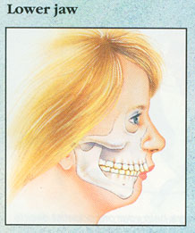  A face that is out of proportion or a bite that is inefficient can be the result of inappropriate growth in either the upper jaw, the lower jaw, or both. Overgrowth of the lower jaw (mandibular hyperplasia) will result in overprojection of the chin and mandible as well as positioning the lower teeth too far forwards, causing an "underbite" or Angle class III malocclusion in technical terms (see leftmost illustration). A face that is out of proportion or a bite that is inefficient can be the result of inappropriate growth in either the upper jaw, the lower jaw, or both. Overgrowth of the lower jaw (mandibular hyperplasia) will result in overprojection of the chin and mandible as well as positioning the lower teeth too far forwards, causing an "underbite" or Angle class III malocclusion in technical terms (see leftmost illustration).
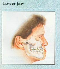
The lower teeth can often be brought into contact with the upper teeth which may make it difficult to bite and chew food. Likewise, the mandible may be underdeveloped (mandibular hypoplasia) resulting in an "overbite" as well as weakness in the projection of the chin and jaw. (see rightmost illustration).
 The facial profile often lacks chin projection and the neck may appear full and not demonstrate the distinct angles associated with a normal mandible. As one might assume, similar development may be disrupted in a purely vertical axis, purely horizontal axis, or any combination thereof. When there is an overdeveloped upper jaw in the vertical axis, the patient may present with a "gummy smile"; all of the teeth as well as a considerable amount of gum may show in a full smile. If the upper jaw is underdeveloped, the middle of the face may look sunken in and the upper teeth will
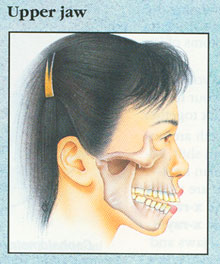
appear receded or may not show at all in the relaxed face. Sometimes only a portion of the jaw is affected; for example the back portion of the upper jaw may alone overdevelop which allows only the back teeth to touch. The front teeth are unable to touch resulting in an open bite. In the illustration at right notice that the front teeth are not in contact resulting in an open bite anteriorly (front). The facial profile often lacks chin projection and the neck may appear full and not demonstrate the distinct angles associated with a normal mandible. As one might assume, similar development may be disrupted in a purely vertical axis, purely horizontal axis, or any combination thereof. When there is an overdeveloped upper jaw in the vertical axis, the patient may present with a "gummy smile"; all of the teeth as well as a considerable amount of gum may show in a full smile. If the upper jaw is underdeveloped, the middle of the face may look sunken in and the upper teeth will
appear receded or may not show at all in the relaxed face. Sometimes only a portion of the jaw is affected; for example the back portion of the upper jaw may alone overdevelop which allows only the back teeth to touch. The front teeth are unable to touch resulting in an open bite. In the illustration at right notice that the front teeth are not in contact resulting in an open bite anteriorly (front).
A Team Approach to Care
The correction of facial imbalance and developmental jaw malpositioning requires a team approach and it is rare that treatment will not involve both an orthodontist and general dentist as well as your oral and maxillofacial surgeon. Although your care may begin with a consultation with your oral and maxillofacial surgeon, the orthodontist will likely initiate therapy. After his exam he will usually obtain records which most often include special radiographs (x-rays) and models fabricated from impressions or molds of your teeth. Your surgeon will also perform a thorough review of your medical history and do his own exam and radiographs as well. Then, after reviewing all of these records together, your doctors can counsel you about your treatment options and help you select the therapy that is best for you.
The orthodontist's role primarily involves positioning the teeth properly within the bone. When jaw growth disturbances are present, our bodies generally try to compensate for them by "growing" the teeth in the direction that tends to compensate for the problem. For example, if a severe overbite is present, the body compensates by erupting the lower teeth out more and trying to "pull" the upper teeth in. These tooth movements tend to compensate or mask the bite disturbance. However, once the jaws have been moved to their appropriate position these compensations will make correcting the final bite terribly difficult. Therefore, an early goal of the orthodontist is to correct these compensations and move the teeth into their appropriate position within the bone of their respective jaw. Now when your jaw is moved into its correct position with surgery the teeth are ready and the bite can be perfected quickly and easily. Because of the early decompensation by the orthodontist, the bite may appear temporarily worse immediately prior to surgery, however, it is important that the teeth be properly positioned in the bone so that the true imbalance can be realized.
The initial examination may also help determine if other conditions need to be managed prior to initiating your therapy. An example would be the presence of wisdom teeth or third molars. The presence of wisdom teeth can complicate both the surgical as well as the orthodontic correction of your bite and will likely need to be removed early in your therapy. The treatment planning stages of your therapy are an ideal time to explore your questions about your treatment course and to mentally prepare yourself for your surgery.
Time Involved
It is important to recognize that surgical/orthodontic treatment is typically not completed quickly. The initial evaluation and treatment planning of a jaw malrelationship involves approximately one to three months. It is during this period that records are taken and evaluated and the orthodontist and surgeon may meet to plan the treatment course. Following this time, we will meet with you and review the available treatment options and help you decide which is right for you.
Since most jaw relationships cannot be corrected without some change in the position of the teeth, it is typically necessary to have six to eighteen months of orthodontic therapy prior to surgical movement of the jaws.
Once the preoperative orthodontic therapy is completed and your orthodontist and surgeon have decided that you are ready for surgery we will arrange a surgery time at your convenience at any of several hospitals or outpatient surgery centers where we operate. The various clerical preparations for your surgery will be made at the hospital. Prior to surgically repositioning the jaws, an exhaustive presurgical workup is performed. New stone models of the bite are made and articulated with special appliances so that the patient's bite can be most accurately duplicated. On these articulated models, the oral surgeon is able to perform planned surgical movements. The entire surgery is first performed on these models in the lab to confirm that the exact jaw movements necessary can be duplicated in the operating room. Custom plastic bite splints are fabricated to ensure that these movements are accurate. If the teeth are adequately aligned and if the surgical movements on the models create a stable bite, the surgery is scheduled. Finally on the morning of your surgery you will meet your anesthesiologist and be prepared for your trip to the operating room. Once there, you will be given medication which allows you to fall asleep, and the surgical team will perform your surgery. If the surgical correction involves moving only one jaw, two to four hours will be needed in the operating room to complete the repositioning. If the correction involves movement of both the upper and lower jaw, six to eight hours in the operating room are required. The postoperative course typically involves an hour in the recovery room, followed by one or two days in the hospital. Following your discharge home, your recovery is typically relatively rapid and uneventful. Although it will depend on the amount of surgery performed, most patients will miss between 6-12 days of work recovering. During the first few weeks after the procedure, the oral surgeon will require several visits to monitor your healing and make sure that your care is proceeding well. After four to six weeks, the visits with the oral surgeon decrease in frequency and the patient returns to the care of the orthodontist. On average, three to ten months of orthodontic therapy are required after surgical repositioning of the jaws. The oral surgeon will also be following the patient during this time to ensure optimal results.
Surgical Procedures
 The vast majority of these surgical procedures are performed intraorally. Incisions are made inside the mouth to access the bony anatomy. Working through these incisions we are able to make the necessary incisions in the bone of the upper and lower jaw. Once the bone segments are free they can be moved to their new position using the plastic splints which were made before the surgery to ensure that the jaw is in the appropriate position. Once we are sure that the position is accurate, fixation appliances are placed to hold the bone rigidly during the healing process. These appliances consist of tiny titanium screws and plates (see right) which rigidly hold the jaw segments where they belong. Once placed the patient will generally NOT need to be wired closed during the healing process. These titanium appliances are quite small and generally imperceptible once placed. The material is extremely safe and well tolerated and therefore, once placed, it is quite rare for removal to be necessary or even desired. The greatest benefits of this technique are that the results of the surgery are more predictable and stable. An additional benefit is that limited jaw function is possible immediately following surgery making eating, talking, and hygiene much easier. It is likely that a small rubber band (elastics) will be placed to help reeducate your muscles following surgery. These will allow you to open and close and can be applied and removed by you for hygiene and eating. It should be pointed out that while this type of fixation will allow jaw function you should not do any chewing following your surgery until specifically told to advance your diet by your surgeon. Until your surgery sites have healed, it is possible to dislodge or move the segments which would likely affect your bite. The vast majority of these surgical procedures are performed intraorally. Incisions are made inside the mouth to access the bony anatomy. Working through these incisions we are able to make the necessary incisions in the bone of the upper and lower jaw. Once the bone segments are free they can be moved to their new position using the plastic splints which were made before the surgery to ensure that the jaw is in the appropriate position. Once we are sure that the position is accurate, fixation appliances are placed to hold the bone rigidly during the healing process. These appliances consist of tiny titanium screws and plates (see right) which rigidly hold the jaw segments where they belong. Once placed the patient will generally NOT need to be wired closed during the healing process. These titanium appliances are quite small and generally imperceptible once placed. The material is extremely safe and well tolerated and therefore, once placed, it is quite rare for removal to be necessary or even desired. The greatest benefits of this technique are that the results of the surgery are more predictable and stable. An additional benefit is that limited jaw function is possible immediately following surgery making eating, talking, and hygiene much easier. It is likely that a small rubber band (elastics) will be placed to help reeducate your muscles following surgery. These will allow you to open and close and can be applied and removed by you for hygiene and eating. It should be pointed out that while this type of fixation will allow jaw function you should not do any chewing following your surgery until specifically told to advance your diet by your surgeon. Until your surgery sites have healed, it is possible to dislodge or move the segments which would likely affect your bite.
If the lower jaw is moved forward, a special separation (osteotomy) is performed (see above right), allowing the bone in which the teeth are housed, to be separated from the bone that is part of the joint complex. The lower jaw is positioned into the new, planned position and screws are placed to secure the two bony segments together.
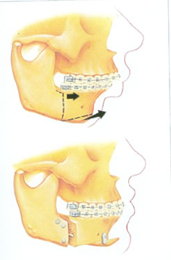
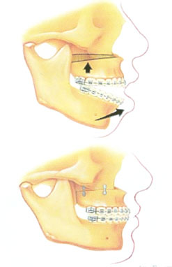 If upper jaw surgery is planned it is likewise performed through incisions inside the mouth. The bone osteotomies are then made and the upper jaw is freed from the rest of the facial skeleton. It can then be moved into its new, correct position as determined by the prefabricated plastic bite splints. Once in place, the segments are fixated with the aforementioned titanium plates and screws to hold it in its proper position during the healing period (see lower right). In the case illustrated at right the maxilla is being moved upwards to correct hyperplasia and, likely, a gummy smile as well as the obvious anterior open bite. Because of the geometry of this movement, some bone will be removed to allow superior movement of the upper jaw, illustrated at right. Again, this surgery typically does NOT require you to be wired closed following the operation and will allow limited jaw function during the healing period.
Recovery
Before and during the hospital stay, information about diet, hygiene, and what to expect will be given to the patient. Because the diet must remain liquid or soft and chewing is discouraged, special dietary arrangements are made to accommodate this. Pamphlets are available to help the patient create varied and balanced meals so that optimal nutrition can be obtained in a non-chew diet. Oral hygiene can be difficult to maintain during the early healing period because of the surgical incisions and swelling. Special techniques and materials will be shared with the patient so that optimal cleanliness can be maintained. Following your discharge from the hospital, most patients will require a week of home recovery. While you are not bedridden during this period of time and most patients are able to engage in normal sedentary activities, you should plan to spend this time in a quiet relaxed environment which will facilitate your recovery.
The inevitable swelling that occurs with surgery peaks at approximately 48 to 72 hours. After this period, the majority of swelling resolves quickly, within one to two weeks. Very subtle changes can occur in the patient's facial presentation over months following the surgical procedure. For most patients, following this week of home convalescence, return to full activity will be made uneventfully.
After the initial healing period of 3 to 6 weeks, you will see your orthodontist to have your progress evaluated. At this time he or she will discuss the timing of your return to active orthodontic therapy and your care will gradually return to purely orthodontic treatment. The period of postoperative orthodontic therapy will vary from patient to patient but is typically between 3 to 9 months. During this period of time the bite will be perfected and final adjustments will be made to the positions of individual teeth. Once this is complete the orthodontic appliances are removed and retainers will be placed to ensure that your results are permanent.
Orthognathic Surgery
 Orthognathic surgery is performed on a regular basis by both Dr. Weil and Dr.
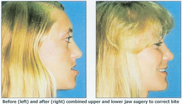
Koo. We would be happy to meet with you to discuss your concerns and answer any questions you might have about surgical orthodontic therapy and the correction of similar bite problems. It should be pointed out that while the correction of malocclusion with orthognathic surgery is best thought of as a functional surgery and happily is generally viewed as medically necessary by insurance providers, there are often wonderful esthetic consequences as a result of these surgeries as well. The patient pictured at right presented with a class III malocclusion (underbite) and was treated with combined upper and lower jaw surgery as well as pre and postoperative orthodontics. One can readily appreciate the dramatic improvement in the facial profile that is a result of this surgery. This sort of esthetic transformation is possible with carefully planned and executed orthognathic surgery. Orthognathic surgery is performed on a regular basis by both Dr. Weil and Dr.
Koo. We would be happy to meet with you to discuss your concerns and answer any questions you might have about surgical orthodontic therapy and the correction of similar bite problems. It should be pointed out that while the correction of malocclusion with orthognathic surgery is best thought of as a functional surgery and happily is generally viewed as medically necessary by insurance providers, there are often wonderful esthetic consequences as a result of these surgeries as well. The patient pictured at right presented with a class III malocclusion (underbite) and was treated with combined upper and lower jaw surgery as well as pre and postoperative orthodontics. One can readily appreciate the dramatic improvement in the facial profile that is a result of this surgery. This sort of esthetic transformation is possible with carefully planned and executed orthognathic surgery.
|